
Radiocarbon dating World Museum's collections

Following news that World Museum could be home to some of the oldest human remains from north-west Europe, Dr Emma Pomeroy explains how radiocarbon dating is helping her research:
Radiocarbon dating involves destroying a tiny piece of the object you want to test. Although this will only leave a small trace on the object itself, it's really important to have a good record of what the teeth and jaw were like. e before they were sampled to preserve them for future research. So on July 8th, we took the teeth and jaw to the Cambridge Biotomography Centre for micro-CT scanning by our colleague, Dr Laura Buck at the University of Cambridge.
 Photo (top left) and 3D models of LIVCM 44.28.WE.3, a lower third molar (wisdom tooth), showing what is possible with the microCT output. Upper right: external surface. Lower left: window cut through surface to show inner structure. Lower right: surfaces made transparent to reveal inner structure.
CT (computed tomography) is the same technology that is used in hospitals to see inside the body. It uses X-ray images taken from many different angles to recreate a 3D version of the object or person. Micro-CT allows very high resolution digital 3D models to be constructed, preserving the teeth and jaws in virtual 3D form for future study. It also enables us to look inside the specimens and see their internal structure. This could be useful for future research, as well as showing whether the teeth have internal damage that we can't see from the outside, such as cracks.
Copies of the digital models will be kept in the Museum as part of their archive. The Museum has also taken photographs of the specimens before they were loaned for sampling.
At Liverpool John Moore’s School of Natural Sciences and Psychology, we recently obtained an Artec Space Spider handheld 3D scanner. While micro-CT gives a fantastic level of internal and external detail, it doesn't preserve the appearance of the objects on the outside - what is known as 'texture' in 3D imaging circles (but essentially what we could call colour). Surface scanners, such as the Artec Space Spider, use structured light to recreate objects in 3D, including their 'texture'. By combining traditional photography with micro-CT scanning and structured light surface scanning, we can create the best possible record of the teeth and jaws in 2D and 3D.
Photo (top left) and 3D models of LIVCM 44.28.WE.3, a lower third molar (wisdom tooth), showing what is possible with the microCT output. Upper right: external surface. Lower left: window cut through surface to show inner structure. Lower right: surfaces made transparent to reveal inner structure.
CT (computed tomography) is the same technology that is used in hospitals to see inside the body. It uses X-ray images taken from many different angles to recreate a 3D version of the object or person. Micro-CT allows very high resolution digital 3D models to be constructed, preserving the teeth and jaws in virtual 3D form for future study. It also enables us to look inside the specimens and see their internal structure. This could be useful for future research, as well as showing whether the teeth have internal damage that we can't see from the outside, such as cracks.
Copies of the digital models will be kept in the Museum as part of their archive. The Museum has also taken photographs of the specimens before they were loaned for sampling.
At Liverpool John Moore’s School of Natural Sciences and Psychology, we recently obtained an Artec Space Spider handheld 3D scanner. While micro-CT gives a fantastic level of internal and external detail, it doesn't preserve the appearance of the objects on the outside - what is known as 'texture' in 3D imaging circles (but essentially what we could call colour). Surface scanners, such as the Artec Space Spider, use structured light to recreate objects in 3D, including their 'texture'. By combining traditional photography with micro-CT scanning and structured light surface scanning, we can create the best possible record of the teeth and jaws in 2D and 3D.
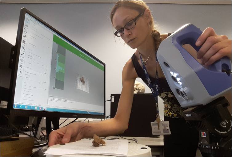 Surface scanning the maxilla (LIVCM 44.28.WD) with the Artec Space Spider at Liverpool John Moores University.